Wood's Lamp Examination: A Comprehensive Guide to Skin Disease Diagnosis
Wood's lamp examination is a non-invasive, convenient, swift, and practical method for detecting skin diseases. It utilizes specific wavelengths of light to cause various fluorescent reactions in skin lesions, aiding in the identification of conditions that are difficult to diagnose with the naked eye. This technique is a valuable supplementary tool in dermatological diagnostics. This consensus elaborates on the Wood's lamp characteristics of several common pigmentary and infectious dermatoses, providing diagnostic and differential diagnostic guidance.
Basic Principle of Wood's Lamp
The Wood's lamp uses a high-pressure mercury lamp as the emission source, producing light at wavelengths of 320-400 nm (peak at 365 nm). This ultraviolet light, when directed at the skin, is scattered or reflected by the epidermis, while pigment precursors, their metabolites, elastic fibers, and collagen fibers in the skin can emit distinct autofluorescence. Consequently, different tissues and pathological changes produce varying fluorescence colors. For optimal examination, the room should be completely dark, and the lamp should be held 10-30 cm away from the lesions.
Characteristics of Pigmentary Disorders Under Wood's Lamp
Melanin has a high capacity to absorb ultraviolet and visible light. When Wood's lamp light is directed at melanin-rich epidermis, it is mostly absorbed, whereas it is scattered and reflected by adjacent melanin-deficient skin, creating a clear demarcation. Wood's lamp examination highlights the contrast between areas of increased or decreased pigmentation and normal skin. In hypopigmented or depigmented lesions, the absence of melanocytes or melanin granules results in fluorescence originating from dermal collagen, appearing as bright blue-white spots with sharp borders. In hyperpigmented lesions, melanin-rich areas absorb more light, appearing black. Notably, epidermal pigment is more visible under Wood's lamp due to its superficial location and fewer interferences, while dermal pigment is less pronounced or unchanged because the surrounding collagen autofluorescence diminishes the contrast.
Wood's Lamp Features of Hypopigmented Diseases
1. Vitiligo: Bright white fluorescence with clear borders.
2. Tuberous Sclerosis: Characteristic ash leaf spots.
3. Ito Hypomelanosis: Swirling or streaky patterns.
4. Nevus Anemicus: Fluorescence not apparent.
5. Achromic Nevus: Grayish-white under Wood's lamp.
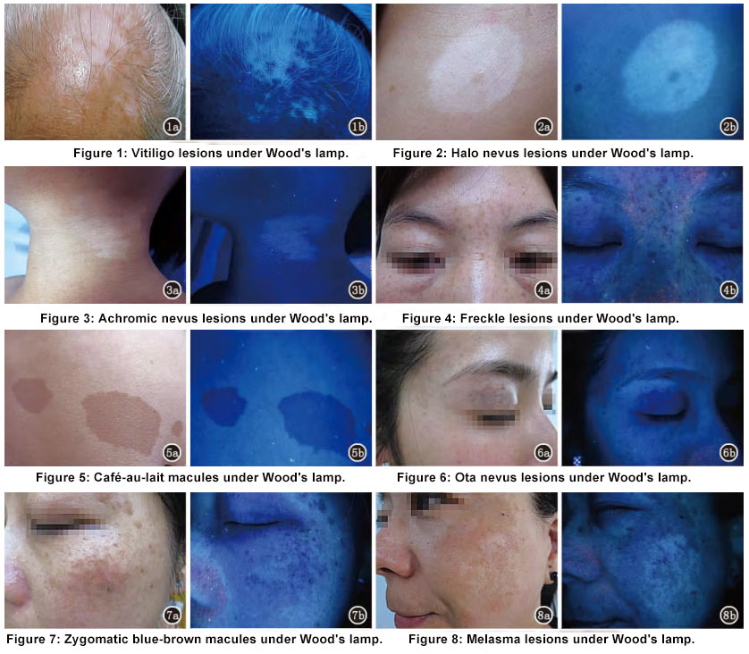
(1) Vitiligo: Under the Wood's lamp, vitiligo lesions show characteristic bright blue-white spots with clear borders. The histopathology of vitiligo reveals a reduction or absence of melanocytes and melanin granules, causing the fluorescence to emanate from dermal collagen, resulting in bright blue-white spots that starkly contrast with surrounding skin. This feature is particularly valuable for early diagnosis and differentiation, especially in patients with fair skin or faint lesions. Wood's lamp can also be used for comprehensive skin screening and examination, often detecting unnoticed lesions.
(2) Halo Nevus: Halo nevus features a central pigmented nevus (usually an intradermal nevus) surrounded by a depigmented halo. Early halo nevus presents as a lighter skin area around the nevus, but under Wood's lamp, the depigmented halo appears blue-white, clearly delineated from the central nevus and adjacent normal skin.
(3) Achromic Nevus: Presenting at birth or shortly after as localized hypopigmented patches. Under Wood's lamp, achromic nevus appears pale blue-white, distinct from the bright blue-white of vitiligo, facilitating differential diagnosis.
Wood's Lamp Features of Hyperpigmented Diseases
(1) Freckles: Due to increased melanin in keratinocytes, freckles appear darker under Wood's lamp as scattered black spots, clearly contrasting with normal skin. Wood's lamp enhances the visibility of freckles compared to natural light, revealing lesions not visible in natural light or those that fade in winter.
(2) Café-au-lait Macules: Increased epidermal melanocytes cause café-au-lait macules to appear as well-defined black-brown patches under Wood's lamp, contrasting sharply with surrounding skin.
(3) Ota Nevus: Similar histopathology to Mongolian spots, with melanocytes scattered in dermal collagen fibers but more superficial. Lesions appear deep blue-brown, clearly contrasting with normal skin.
(4) Zygomatic Blue-Brown Macules: Present as gray-brown, gray-blue, or deep brown round or oval spots on the zygomatic region, symmetrically distributed. Under Wood's lamp, these lesions appear blue-black, clearly contrasting with normal skin.
(5) Melasma: Under Wood's lamp, melasma appears as blue-black patches with clear borders. Sanchez et al. reported using Wood's lamp to classify melasma. Epidermal melasma darkens under Wood's lamp compared to visible light; dermal melasma appears pale blue under natural light and does not darken under Wood's lamp. This classification aids in determining treatment and prognosis, with epidermal melasma responding better to bleaching agents and other topical treatments. Wood's lamp serves as an effective method for monitoring treatment and prognosis.

Wood's Lamp Features of Infectious Skin Diseases
Certain pathogens fluoresce under Wood's lamp, aiding in diagnosing infectious diseases.
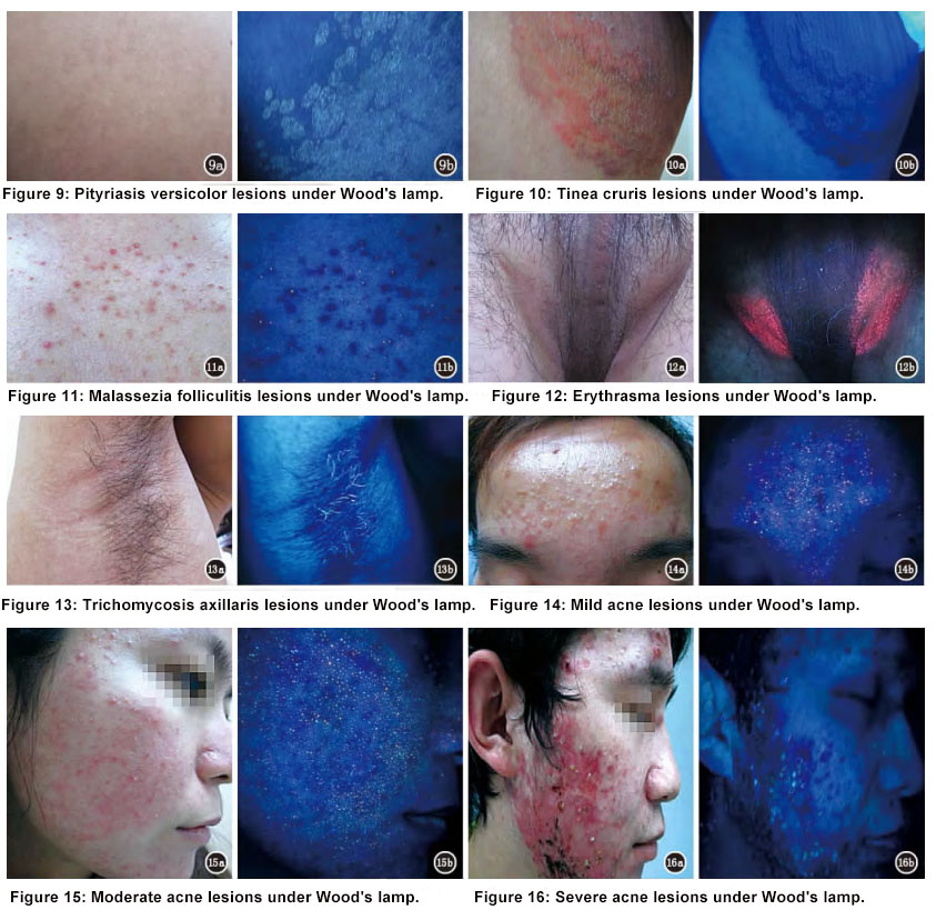
(1) Pityriasis Versicolor: Caused by Malassezia yeast, appearing as yellow-green or yellow fluorescence under Wood's lamp.
(2) Tinea Capitis: The first application of Wood's lamp in dermatology. Caused by various dermatophytes, with fluorescence features listed in Table 2
Table 2. Wood's Lamp Fluorescence Characteristics of Tinea Capitis
| Pathogen | Fluorescence Color |
| Microsporum audouinii | Blue-green |
| Microsporum canis | Blue-green |
| Microsporum ferrugineum | Blue-green |
| Microsporum distortum | Blue-green |
| Microsporum gypseum | Dark yellow |
| Trichophyton schoenleinii | Dark blue |
| Trichophyton violaceum | No fluorescence |
| Trichophyton tonsurans | No fluorescence |
| Trichophyton verrucosum | No fluorescence |
(3) Tinea Cruris: Fungal infection of the groin, perineum, perianal area, and buttocks, caused by Trichophyton rubrum, Epidermophyton floccosum, etc. Most show clear ring-like blue-black patches under Wood's lamp, without fluorescence.
(4) Malassezia Folliculitis: Caused by Malassezia, affecting young adults, mainly on the chest and back. Typical lesions are inflammatory follicular papules or pustules, 2-4 mm in diameter, densely or sparsely distributed. Blue-black patches with some showing blue-white pinpoint fluorescence aid in differential diagnosis.
Bacterial Infectious Skin Diseases
(1) Erythrasma: Caused by Corynebacterium minutissimum, presenting as coral red fluorescence under Wood's lamp, distinguishing it from tinea cruris.
(2) Trichomycosis Axillaris: Caused by Corynebacterium, infecting axillary and pubic hair, appearing as bright blue-white fluorescence under Wood's lamp.
(3) Pseudomonas Infections: Early detection of Pseudomonas infections, especially in burn wounds, is possible with Wood's lamp. Pathogens produce pyoverdine, visible as green fluorescence. Fluorescence indicates potential infection, requiring immediate treatment.
(4) Acne: Caused by Propionibacterium acnes, producing coproporphyrin, visible as orange-red fluorescence under Wood's lamp. Fluorescence correlates with P. acnes density. Severe acne with nodules, cysts, and scars shows less or no fluorescence.

